热带病与寄生虫学 ›› 2026, Vol. 24 ›› Issue (1): 47-53.doi: 10.20199/j.issn.1672-2302.2026.01.009
胡婷婷1( ), 赵成思2(), 林树晴1, 王桂芳1, 邱竞帆1, 张戎1, 刘新建1(), 王勇1()
收稿日期:2025-10-24
出版日期:2026-02-20
发布日期:2026-03-31
通信作者:
刘新建,E-mail: 作者简介:胡婷婷,女,硕士在读,研究方向:感染与免疫。E-mail: 基金资助:
HU Tingting1(), ZHAO Chengsi2(), LIN Shuqing1, WANG Guifang1, QIU Jingfan1, ZHANG Rong1, LIU Xinjian1(), WANG Yong1()
Received:2025-10-24
Online:2026-02-20
Published:2026-03-31
Contact:
LIU Xinjian, E-mail: 摘要:
目的 探讨日本血吸虫慢性感染对小鼠肾脏纤维化的动态影响及其分子机制,为阐明血吸虫病肾病的发病机制提供实验依据。方法 建立C57BL/6J小鼠日本血吸虫慢性感染的模型(15只),同时设置未感染组(15只)。分别于感染后第12周、14周、16周收集小鼠肾脏组织(感染组和未感染组各5只)进行检测分析。采用qRT-PCR检测肾脏组织中纤维化标志物(α-SMA、CTGF、Col-1、Col-4)的mRNA表达水平,Western blot检测CTGF、Col-1、P-Smad2/3及Smad7的蛋白表达,免疫组织化学检测Smad7蛋白的表达丰度与细胞定位。使用日本血吸虫可溶性成虫抗原(soluble worm antigen, SWA)与转化生长因子-β(transforming growth factor-β, TGF-β)信号通路激活剂TGF-β1刺激体外培养的小鼠肾小球系膜细胞(MES-13),通过qRT-PCR检测纤维化标志物、Smad3和Smad7的mRNA表达水平,免疫荧光观察Col-1和α-SMA蛋白在细胞中的表达情况。结果 qRT-PCR结果显示,相较于未感染组,12周、14周、16周感染组小鼠肾脏组织α-SMA、CTGF、Col-1及Col-4 mRNA表达均上调(t=4.61、6.64、3.52,t=3.29、5.07、7.22,t=3.66、4.74、3.10,t=3.24、5.92、2.67;P均<0.05)。Western blot结果显示,相较于未感染组,各时间点感染组CTGF、Col-1蛋白表达均上调(t=4.07、7.39、8.84,t=3.08、4.21、4.85;P均<0.05);14周感染组肾脏组织P-Smad2/3蛋白表达高于未感染组(t=3.61,P<0.05),Smad7蛋白表达低于未感染组(t=7.96,P<0.05)。体外实验表明,SWA与TGF-β1干预后,MES-13细胞α-SMA、CTGF、Col-1、Col-4及Smad3 mRNA表达均高于未干预组,Smad7 mRNA表达低于未干预组,3组mRNA表达差异有统计学意义(F=62.26、112.70、7.64、127.20、10.78、6.75,P均<0.05);免疫荧光检测证实SWA组α-SMA与Col-1蛋白表达增强。结论 日本血吸虫慢性感染期小鼠肾脏存在持续纤维化状态,SWA直接激活TGF-β信号通路在纤维化致病中可能起到重要作用。
中图分类号:
胡婷婷, 赵成思, 林树晴, 王桂芳, 邱竞帆, 张戎, 刘新建, 王勇. 日本血吸虫慢性感染诱发小鼠肾脏纤维化的实验研究[J]. 热带病与寄生虫学, 2026, 24(1): 47-53.
HU Tingting, ZHAO Chengsi, LIN Shuqing, WANG Guifang, QIU Jingfan, ZHANG Rong, LIU Xinjian, WANG Yong. Experimental study on renal fibrosis induced by chronic Schistosoma japonicum infection in mouse models[J]. Journal of Tropical Diseases and Parasitology, 2026, 24(1): 47-53.
表1
qRT-PCR检测引物序列
| 基因名称 | 上游引物序列(5'-3') | 下游引物序列(5'-3') |
|---|---|---|
| GAPDH | TGGATTTGGACGCATTGGTC | TTTGCACTGGTACGTGTTGAT |
| CTGF | AACAGTGGAGATGCCAGGAG | TAATTTCCCTCCCCGGTTAC |
| Col-1 | GTCGAGGGCCAAGACGAAG | CAGATCACGTCATCGCACAAC |
| α-SMA | CAGGGCTGTTTTCCCATCCAT | GCCATGTTCTATCGGGTACTTC |
| Col-4 | TTCAGATTCCGCAGTGCCCTA | TTCTCATGCACACTTGGCAGC |
| Smad3 | CCATCTCCTACTACGAGCTGAA | CACTGCTGCATTCCTGTTGAC |
| Smad7 | GTGTTGCTGTGAATCTTACG | AGAAGAAGTTGGGAATCTGA |
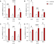
图1
日本血吸虫慢性感染对小鼠肾脏纤维化相关指标的基因水平表达的影响 注:A为α-SMA mRNA表达水平;B为CTGF mRNA表达水平;C为Col-1 mRNA表达水平;D为Col-4 mRNA表达水平;*表示P<0.05;**表示P<0.01;***表示P<0.001。


图2
日本血吸虫慢性感染对小鼠肾脏纤维化相关指标的蛋白表达水平的影响 注:A、C为CTGF蛋白表达水平;B、D为Col-1蛋白表达水平;*表示P<0.05;**表示P<0.01。
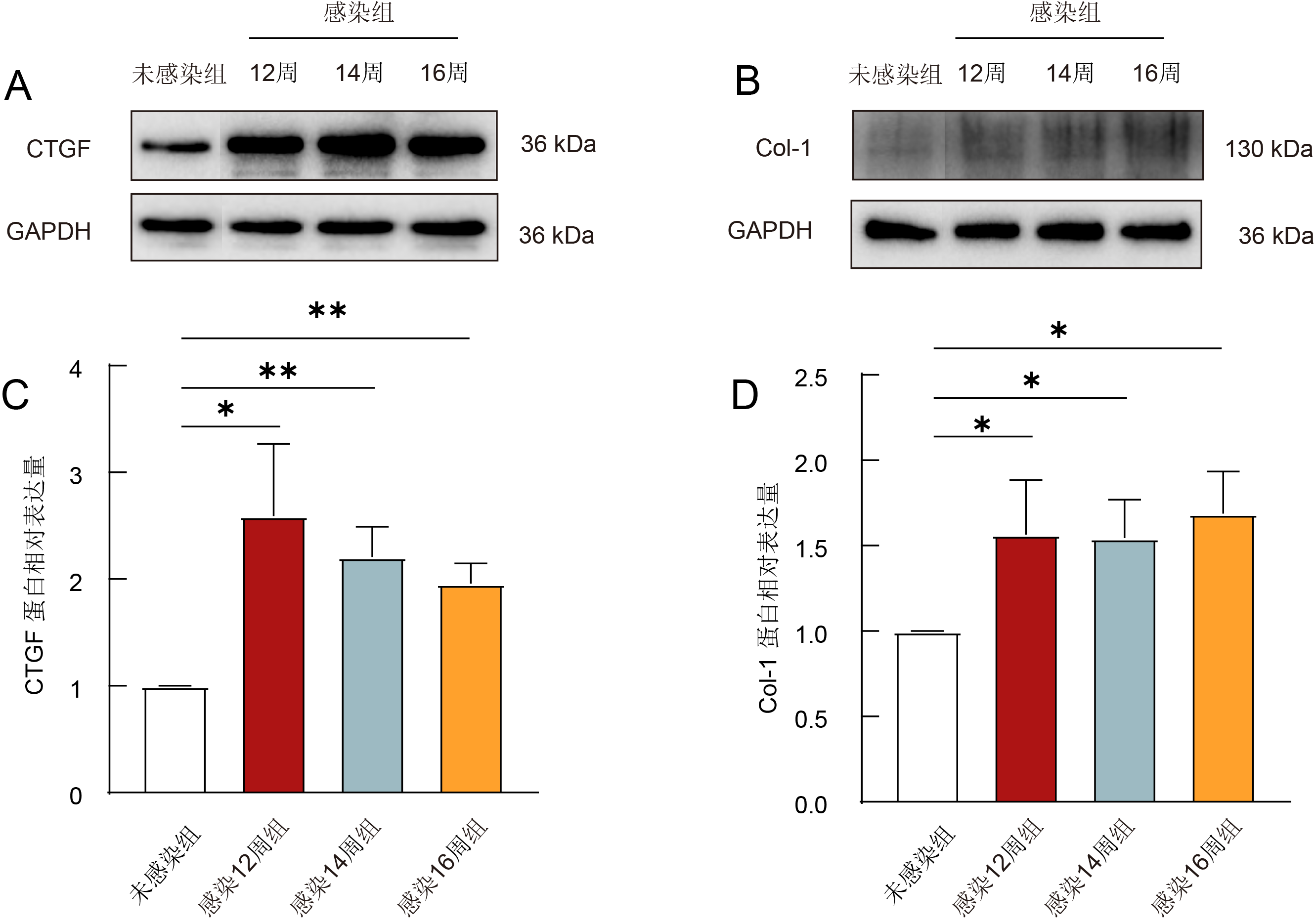

图3
日本血吸虫慢性感染对小鼠肾脏TGF-β信号通路相关指标的基因表达水平的影响 注:A为Smad3 mRNA表达水平;B为Smad7 mRNA表达水平;**表示P<0.01;***表示P<0.001。
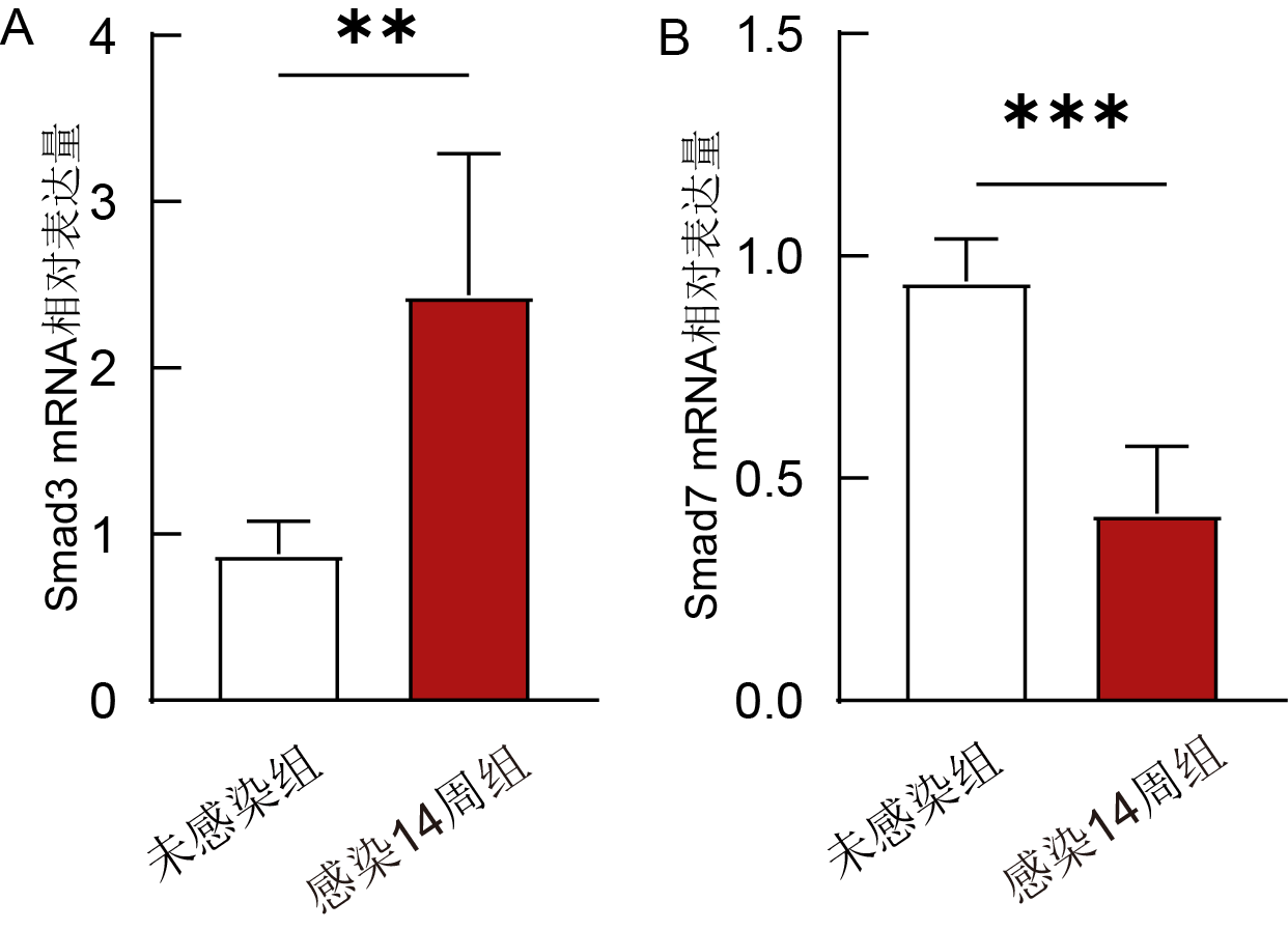
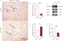
图4
日本血吸虫慢性感染对小鼠肾脏TGF/β信号通路相关指标的蛋白表达水平的影响 注:A为两组小鼠肾脏组织中Smad7阳性细胞的浸润情况,箭头指示典型的Smad7阳性区域;B为两组小鼠肾脏组织Smad7阳性区域表达水平;C-E为两组小鼠肾脏组织P-Smad2/3、Smad7蛋白表达水平;*表示P<0.05;**表示P<0.01。

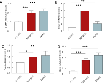
图5
SWA刺激对MES-13细胞纤维化相关指标基因表达水平的影响 注:A为α-SMA基因表达水平;B为CTGF基因表达水平;C为Col-1基因表达水平;D为Col-4基因表达水平;*表示P<0.05;**表示P<0.01;***表示P<0.001。

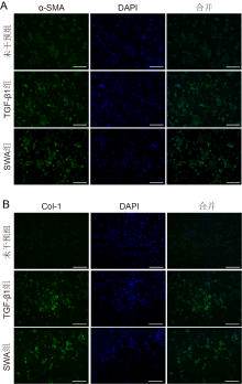
图6
SWA刺激对MES-13细胞纤维化相关指标蛋白表达水平的影响 注:A为α-SMA蛋白表达水平;B为Col-1蛋白表达水平。
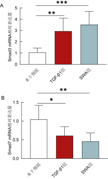
图7
SWA刺激对MES-13细胞TGF-β相关指标基因表达水平的影响 注:A为Smad3基因表达水平;B为Smad7基因表达水平;*表示P<0.05;**表示P<0.01;***表示P<0.001。

| [1] |
Zhou Y, Zheng M, Gong YF, et al. Changing seroprevalence of schistosomiasis Japonica in China from 1982 to 2020: a systematic review and spatial analysis[J]. PLoS Negl Trop Dis, 2024, 18(9):e0012466.
doi: 10.1371/journal.pntd.0012466 URL |
| [2] |
Wilson MS, Mentink-Kane MM, Pesce JT, et al. Immunopathology of schistosomiasis[J]. Immunol Cell Biol, 2007, 85(2):148-154.
doi: 10.1038/sj.icb.7100014 pmid: 17160074 |
| [3] |
de Menezes Neves PDM, Jorge LB, Cavalcante LB, et al. Schistosomiasis-associated glomerulopathy: Clinical aspects, pathological characteristics, and renal outcomes[J]. Clin Nephrol, 2020, 93(5):251-261.
doi: 10.5414/CN110013 URL |
| [4] |
Liao ZN, Tao LJ, Yin HL, et al. Schistosoma japonicum infection associated with membranous nephropathy: a case report[J]. BMC Infect Dis, 2022, 22(1):125.
doi: 10.1186/s12879-022-07092-0 |
| [5] |
Houba V. Experimental renal disease due to schistosomiasis[J]. Kidney Int, 1979, 16(1):30-43.
pmid: 119088 |
| [6] |
赵成思, 秦敏, 谭明娟,等. 吡喹酮对日本血吸虫急性感染小鼠肾脏功能损伤的影响[J]. 中国寄生虫学与寄生虫病杂志, 2021, 39(2):200-209.
doi: 10.12140/j.issn.1000-7423.2021.02.013 |
| [7] |
Liu ZL, Zhang LC, Liang YM, et al. Pathology and molecular mechanisms of Schistosoma japonicum-associated liver fibrosis[J]. Front Cell Infect Microbiol, 2022, 12:1035765.
doi: 10.3389/fcimb.2022.1035765 URL |
| [8] |
Hu HH, Chen DQ, Wang YN, et al. New insights into TGF-β/Smad signaling in tissue fibrosis[J]. Chem Biol Interact, 2018, 292:76-83.
doi: 10.1016/j.cbi.2018.07.008 URL |
| [9] |
Lathan R. Exploring unconventional targets in myofibroblast transdifferentiation outside classical TGF-β signaling in renal fibrosis[J]. Front Physiol, 2024, 15:1296504.
doi: 10.3389/fphys.2024.1296504 URL |
| [10] |
Feng LX, Chen C, Xiong X, et al. PS-MPs promotes the progression of inflammation and fibrosis in diabetic nephropathy through NLRP3/Caspase-1 and TGF-β1/Smad2/3 signaling pathways[J]. Ecotoxicol Environ Saf, 2024, 273:116102.
doi: 10.1016/j.ecoenv.2024.116102 URL |
| [11] |
Schwalm S, Beyer S, Frey H, et al. Sphingosine kinase-2 deficiency ameliorates kidney fibrosis by up-regulating Smad7 in a mouse model of unilateral ureteral obstruction[J]. Am J Pathol, 2017, 187(11):2413-2429.
doi: S0002-9440(17)30317-6 pmid: 28807595 |
| [12] |
Hong Q, Kim H, Cai GY, et al. Modulation of TGF-β signaling new approaches toward kidney disease and fibrosis therapy[J]. Int J Biol Sci, 2025, 21(4):1649-1665.
doi: 10.7150/ijbs.101548 pmid: 39990662 |
| [13] |
de Freitas Galvão RL, Meneses GC, Pinheiro MCC, et al. Kidney injury biomarkers and parasitic loads of Schistosoma mansoni in a highly endemic area in northeastern Brazil[J]. Acta Trop, 2022, 228:106311.
doi: 10.1016/j.actatropica.2022.106311 URL |
| [14] |
van Velthuysen ML, Florquin S. Glomerulopathy associated with parasitic infections[J]. Clin Microbiol Rev, 2000, 13(1):55-66.
doi: 10.1128/CMR.13.1.55 pmid: 10627491 |
| [15] |
Zhang WJ, Chen SJ, Zhou SC, et al. Inflammasomes and fibrosis[J]. Front Immunol, 2021, 12:643149.
doi: 10.3389/fimmu.2021.643149 URL |
| [16] |
Cui J, Hong PP, Li ZZ, et al. Chloroquine inhibits NLRP3 inflammasomes activation and alleviates renal fibrosis in mouse model of hyperuricemic nephropathy with aggravation by a high-fat-diet[J]. Int Immunopharmacol, 2023, 120:110353.
doi: 10.1016/j.intimp.2023.110353 URL |
| [17] |
Liao Y, Tan RZ, Li JC, et al. Isoliquiritigenin attenuates UUO-induced renal inflammation and fibrosis by inhibiting mincle/syk/NF-kappa B signaling pathway[J]. Drug Des Devel Ther, 2020, 14:1455-1468.
doi: 10.2147/DDDT.S243420 URL |
| [18] |
He Y, Deng B, Liu SL, et al. Myeloid Piezo1 deletion protects renal fibrosis by restraining macrophage infiltration and activation[J]. Hypertension, 2022, 79(5):918-931.
doi: 10.1161/HYPERTENSIONAHA.121.18750 URL |
| [19] |
Di XP, Li Y, Wei JW, et al. Targeting fibrosis: from molecular mechanisms to advanced therapies[J]. Adv Sci, 2025, 12(3):2410416.
doi: 10.1002/advs.v12.3 URL |
| [20] |
Li L, Fu HY, Liu YH. The fibrogenic niche in kidney fibrosis: components and mechanisms[J]. Nat Rev Nephrol, 2022, 18(9):545-557.
doi: 10.1038/s41581-022-00590-z pmid: 35788561 |
| [21] |
Toda N, Mukoyama M, Yanagita M, et al. CTGF in kidney fibrosis and glomerulonephritis[J]. Inflamm Regen, 2018, 38:14.
doi: 10.1186/s41232-018-0070-0 pmid: 30123390 |
| [22] |
Zhao X, Kwan JYY, Yip K, et al. Targeting metabolic dysregulation for fibrosis therapy[J]. Nat Rev Drug Discov, 2020, 19(1):57-75.
doi: 10.1038/s41573-019-0040-5 pmid: 31548636 |
| [23] |
El Karoui K, Fervenza FC, De Vriese AS. Treatment of IgA nephropathy: a rapidly evolving field[J]. J Am Soc Nephrol, 2024, 35(1):103-116.
doi: 10.1681/ASN.0000000000000242 URL |
| [24] |
Dhakal AK, Shrestha D, Preston R, et al. Acute post-streptococcal glomerulonephritis in children-treatment standard[J]. Nephrol Dial Transplant, 2025, 40(10):1843-1853.
doi: 10.1093/ndt/gfaf130 URL |
| [25] |
Yashima A, Mizuno M, Yuzawa Y, et al. Mesangial proliferative glomerulonephritis in murine malaria parasite, Plasmodium chabaudi AS, infected NC mice[J]. Clin Exp Nephrol, 2017, 21(4):589-596.
doi: 10.1007/s10157-016-1339-8 URL |
| [26] |
Qi X, Pu YN, Chen FY, et al. Schistosome egg antigen stimulates the secretion of miR-33-carrying extracellular vesicles from macrophages to promote hepatic stellate cell activation and liver fibrosis in schistosomiasis[J]. PLoS Negl Trop Dis, 2023, 17(5):e0011385.
doi: 10.1371/journal.pntd.0011385 URL |
| [1] | 马晓荷, 章乐生, 汪峰峰, 路标, 孙成松, 李清越, 王旗, 操治国, 汪天平. 基于深度学习技术的日本血吸虫抗体检测结果智能判读模型的建立和效能评价[J]. 热带病与寄生虫学, 2024, 22(6): 358-363. |
| [2] | 王旗, 章乐生, 汪峰峰, 汪敏, 王毓洁, 马晓荷, 李清越, 操治国. 日本血吸虫成虫和虫卵排泄分泌抗原对Ⅰ型糖尿病模型小鼠的影响[J]. 热带病与寄生虫学, 2024, 22(4): 239-243. |
| [3] | 李宗光, 何婷婷, 谢婧姿, 吕尚标, 胡飞, 袁敏, 林丹丹, 李宜锋. 江西省血吸虫性肝纤维化人群流行病学特征分析[J]. 热带病与寄生虫学, 2024, 22(1): 31-36. |
| [4] | 马晓荷, 汪敏, 朱磊, 郭见多, 李清越, 刘婷, 翟杜娟, 孙成松, 张世清, 汪天平. 安徽省不同地区日本血吸虫群体线粒体基因遗传变异研究[J]. 热带病与寄生虫学, 2021, 19(5): 254-. |
| [5] | 金郁, 刘道华, 金伟, 呼明闯, 汪奇志. IL-10 调节日本血吸虫感染小鼠肝脏炎症和纤维化的实验观察[J]. 热带病与寄生虫学, 2021, 19(5): 259-. |
| [6] | 陈舒心, 秦铭, 周方斌, 何兴. 日本血吸虫与曼氏血吸虫的致病差异[J]. 热带病与寄生虫学, 2021, 19(2): 112-115,封三. |
| [7] | 詹惕, 崔代文, 项可霞, 汪为春, 朱应富, 段永梅. 安徽马鞍山市博望区“有螺无病”成因调查分析 [J]. 热带病与寄生虫学, 2020, 18(2): 115-117. |
| [8] | 张世清, 章乐生, 汪峰峰, 汪天平 . 《日本血吸虫抗体检测 间接红细胞凝集试验》 (WS / T 630-2018)标准解读[J]. 热带病与寄生虫学, 2020, 18(1): 1-4. |
| [9] | 徐桂娜 何雪梅 周晓蓉 曾凡胜 秦志强. 日本血吸虫虫卵分泌物小 RNA的高通量测序[J]. 热带病与寄生虫学, 2019, 17(4): 210-213. |
| [10] | 卢美 邹翔 万圣 杨瑞军 杜海娟 张剑锋. 浙江开化县应用LAMP技术检测感染性钉螺的效果分析[J]. 热带病与寄生虫学, 2019, 17(4): 229-232. |
| [11] | 田添,王培,吕超,秦志强. 日本血吸虫脂筏蛋白的重组表达及其生物信息学分析[J]. 热带病与寄生虫学, 2018, 16(2): 67-. |
| [12] | 曾凡胜,何露,何雪梅,杨杰,秦志强. 日本血吸虫巢式PCR检测方法的建立[J]. 热带病与寄生虫学, 2017, 15(3): 136-. |
| [13] | 何家昶,张世清,汪天平,汪峰峰,尹晓梅,周莉. IHA筛查结果作为血吸虫病疫情判定指标应用价值探讨[J]. 热带病与寄生虫学, 2017, 15(2): 96-98. |
| [14] | 孙成松,朱海,王玥,汪峰峰,尹晓梅,周莉,汪奇志,张世清,汪天平*. 小鼠感染日本血吸虫尾蚴时龄与粪便虫卵排出关系的研究[J]. 热带病与寄生虫学, 2017, 15(1): 19-21. |
| [15] | 喻祎哲,杨杰,曾凡胜,王红梅,秦志强*. 日本血吸虫成虫的非编码RNA高通量测序分析[J]. 热带病与寄生虫学, 2017, 15(1): 31-35. |
| 阅读次数 | ||||||
|
全文 |
|
|||||
|
摘要 |
|
|||||